Virtual Library
Start Your Search
Hiroshi Yoshida
Author of
-
+
MA15 - Usage of Computer and Molecular Analysis in Treatment Selection and Disease Prognostication (ID 141)
- Event: WCLC 2019
- Type: Mini Oral Session
- Track: Pathology
- Presentations: 1
- Now Available
- Moderators:John Le Quesne, Noriko Motoi
- Coordinates: 9/09/2019, 15:45 - 17:15, Tokyo (1982)
-
+
MA15.03 - Exploring Digital Pathology-Based Morphological Biomarkers for a Better Patients’ Selection to the Immune Checkpoint Inhibitor of Lung Cancer (Now Available) (ID 1777)
15:45 - 17:15 | Author(s): Hiroshi Yoshida
- Abstract
- Presentation
Background
For eligible patients’ selection for immune checkpoint inhibitor therapy (ICI), it is important to establish more accurate predicting biomarkers, in addition to PD-L1 IHC and MSI-high. We hypothesized that morphological characteristics should reflect genetic alteration, thus could predict ICI responsiveness. In this study, we examined the predictive potential of morphological characteristics using digital whole-slide images as a new biomarker for ICI-treatment on non-small cell lung cancer (NSCLC) and their relationship to PD-L1 IHC and genetic alterations.
Method
71 NSCLC who received ICI therapy were recruited. Digital images of H&E and PD-L1 (22C3) IHC stained slides of pre-treatment biopsied or resected materials were examined by previously reported image analysis techniques using e-Pathologist ® (NEC, Japan). Morphological characteristics of cancer cells (three and six parameters of nuclear shape and chromatin texture) were extracted as MC-scores. Of 11 cases (pilot cohort), PD-L1 IHC (22C3) and tumor mutation burden (TMB) by the NGS-based target sequence (NCC oncopanel ®) were examined. Correlation between MC-score, PD-L1 IHC, TMB status, and clinical outcome was calculated. A p-value of less than 0.05 was defined as statistically significant.
Decision tree analysis for evaluating predicting ICI-responsiveness was built using statistically significant MC-scores. We also tested the predictive value of a deep learning analysis (AI model) with 5-fold cross-validation. AUC (area under the curve) of ROC analysis was calculated.
Result
Of the responders, the MC-score of cancer cell were statistically different from those of the non-responders; nuclear texture contour complexity (11.8 vs. 8.25, median value of responder vs. non-responders; p<0.01), homogeneity (0.396 vs. 0.421; p<0.01), angular second moment (ASM) (0.0203 vs. 0.0214; p=0.049) and nuclear circularity (0.878 vs. 0.885, p=0.026). Circularity (p=0.011) and texture homogeneity (p=0.048) correlated with TMB. ASM texture correlated with PD-L1 expression (p=0.018). The decision tree model for predictive and screening purposes resulted in 0.83 and 0.62 accuracies, respectively. AUC of AI-model for ICI responsiveness resulted in fair (0.74 on average, range 0.55-0.81).
Conclusion
Our results indicate the substantial value of the morphological feature as a biomarker for ICI therapy. Morphological characteristics are eligible from archived FFPE samples, showed good correlation to the underlying genetic alteration. Digital pathology can serve useful predictive morphological biomarkers for precision medicine of lung cancer patients, and promising the power of AI-assisted pathology.
Only Members that have purchased this event or have registered via an access code will be able to view this content. To view this presentation, please login, select "Add to Cart" and proceed to checkout. If you would like to become a member of IASLC, please click here.
-
+
P1.09 - Pathology (ID 173)
- Event: WCLC 2019
- Type: Poster Viewing in the Exhibit Hall
- Track: Pathology
- Presentations: 1
- Moderators:
- Coordinates: 9/08/2019, 09:45 - 18:00, Exhibit Hall
-
+
P1.09-11 - Influences of Sampling Method to Morphological Feature Measurement of Lung Cancer Cell (ID 886)
09:45 - 18:00 | Author(s): Hiroshi Yoshida
- Abstract
Background
Progress of imaging technologies in the field of histopathology enables us to exploit artificial intelligence (AI) techniques to detect cancer based on digital images for screening or quality assurance of diagnosis process. Nowadays, reports on the application of AI to cancer detection which claim 99-percent detection accuracy are found in every proceedings or journal of digital pathology. However, little attention has been paid to the influences of sampling method to AI-based histological diagnosis.
Method
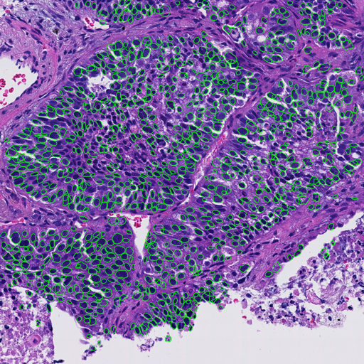
Whole slide images of hematoxylin and eosin (H&E) stained slides collected from 94 non-small cell lung cancer (NSCLC) cases were captured by a virtual slide scanner (NanoZoomer, Hamamatsu Photonics, Japan). Sampling methods were needle biopsy (59 cases), operation (12 cases) and endobronchial ultrasound-guided transbronchial needle aspiration (EBUS-TBNA) (29 cases). Regions of interest (ROI) were selected by an experienced pathologist. After selecting tumor cells only by AI-based tumor cell detecter (Figure.1), following morphological features were calculated: nuclear area, perimeter (Peri), circularity (Circ) and five texture features, i.e., angular secondary moment (ASM), contrast(Cont), homogeneity (Hom) and entropy (Ent) of gray level co-occurrence matrix (GLCM), and contour complexity (CC).
Result
We found significant differences (p<0.05) in most of feature values except nuclear area and perimeter.
Conclusion
Our results suggest that methods of sampling significantly affect morphological feature values of nucleus and this fact must be taken into consideration when applying AI-based techniques to tissue image classification.


