Virtual Library
Start Your Search
Xiang Li
Author of
-
+
OA10 - Sophisticated TNM Staging System for Lung Cancer (ID 136)
- Event: WCLC 2019
- Type: Oral Session
- Track: Staging
- Presentations: 1
- Now Available
- Moderators:Ke-Neng Chen, Pedro Lopez De Castro
- Coordinates: 9/09/2019, 14:00 - 15:30, Toronto (1985)
-
+
OA10.06 - Transition Patterns Between N1 and N2 Stations Discovered from Data-Driven Lymphatic Metastasis Study in Non-Small Cell Lung Cancer (Now Available) (ID 1499)
14:00 - 15:30 | Author(s): Xiang Li
- Abstract
- Presentation
Background
N staging process was essential for evaluation of outcome and indication of following adjuvant therapies in Non-small-cell lung cancer treatment. Various clinical observations on the potential transition patterns of lymph node drainage are reported, however, most of the previous conclusions were made by clinical physicians and focused on specific empirical transition patterns. The fact that there is no definitive and holistic map for lymphatic metastasis transition patterns, and the patients were suffering from either excessive nodes collection along with more damage, or insufficient nodes collection with potential recurrent risks.
Method
We perform complete lymph node examination of a total of 936 subjects diagnosed with NSCLC lung cancer. Lymph nodes sampling or dissection are performed according to NCCN guidelines.
A probabilistic model is developed due to the presence of these missing values. Using the maximum likelihood estimation and proximal gradient algorithm, the summarization of dataset is obtained, which were several explicit metastases and their corresponding probabilities. The metastasis graph is constructed from the summarization result with greedy algorithm and a given threshold. Besides, numerical simulation experiments are conducted to validate the stability of algorithms.
Result
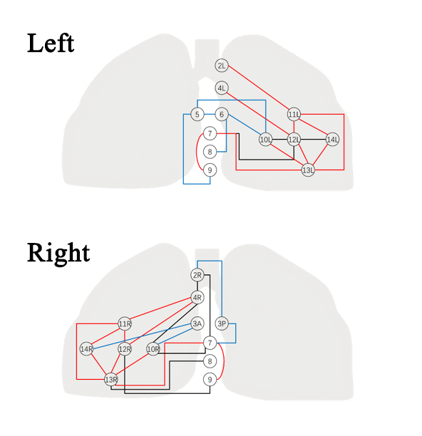
Lymph node sites are shown as round circles according to their anatomical locations. The inferred transition paths are shown as edges connecting them. Edges colored in red are those consistently found in the left and right lung, and blue for unique nodes at each side thus cannot be compared, and black for different patterns between left and right lungs.
Closely connected intra-lobar (N1) nodes: strong connections among intra-lobar nodes (10~14). Over 78% among all the patients have more than 7 edges connecting the 5 intra-lobar nodes.
Jumping metastasis from N1 to N2 stations: We found that there exists several jumping metastasis at both sides of the lobes which has not been well-studied yet posing a challenge for the diagnosis and accurate staging (eg. 12 to 4, 13 to7, 11 to 2), revealing potential long-range transition pathways.
Correlation among N2 lymph nodes: We discovered the presence of certain metastatic groups, including node 5/6, 5/9, 7/9, 6/8 for left lung, and 2/4,2/7,7/9, 2/3p, 3p/7 for right lung.
Conclusion
So we drew a map precisely to make a better understanding for metastatic pathways and provide a potential tool for the prediction of involved nodes pre/ or intro-operatively,so that an individualized surgical planning strategy could be made.
Only Members that have purchased this event or have registered via an access code will be able to view this content. To view this presentation, please login, select "Add to Cart" and proceed to checkout. If you would like to become a member of IASLC, please click here.


